Up-Date/Most Recent

Showing posts with label Metastasize. Show all posts

Brain Metastasis

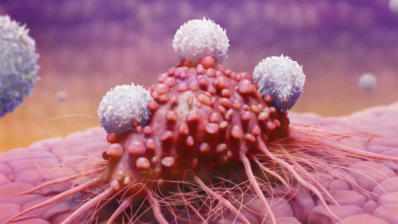
Research conducted at the University of Oxford and published in the journal PLoS One, has succeeded in identifying the mechanism by which cancer cells can enter the blood vessels to the brain. This will be useful for making new types of drugs with mechanisms that prevent cancer cells from spreading and growing in the brain.

Metastasis is the process of spreading and growing cancer cells from the main cancer mass to the surrounding organs. This is the main reason why cancer is a dangerous disease and causes death.

Metastases to the brain are the most common cause of cancer that develops in the brain or central nervous system. The number of brain metastases is up to 10 times that of cancer originating from the brain itself (original). Once cancer cells metastasize to the brain, the patient's prognosis in the future is not very good. Generally, patients can only survive 9 months with maximum therapy. About 20% of all cancer patients will metastasize to the brain.

"Metastasis to the brain is a terminal stage of cancer and very little is known about this process," said dr. W Shawn Carbonell, a postdoctoral researcher at MRC/CRUK Gray Institute for Radiation Oncology and Biology, University of Oxford. "However, by conducting various studies on this matter, it is hoped that we will be able to provide a better therapy to overcome the spread of this cancer."

A research team from the University of Oxford led by professor Ruth Muschel from the Gray Institute for Radiation Oncology and Biology tried to answer the question: how do cancer cells grow and develop in the brain? They studied various types of cancer cells from both humans and mice (breast cancer cells, melanoma, lymphoma) and examined how these cancer cells grow in the brain (in vitro).

The researchers found that cancer cells started growing through the walls of the brain's blood vessels in 95% of cases, rather than in the nerve cells directly. Researchers suspect that by controlling the blood vessels in the brain, cancer cells will obtain nutrients and oxygen and can reproduce without the need to grow their own blood vessels. In addition, cancer cells need brain blood vessels to invade the whole brain.

The dependence of cancer cells on blood vessels in the brain is an opening to help find new therapies for cancer, said professor Muschel. This discovery is one part of the puzzle where it is a breakthrough to find a therapy to prevent the spread of cancer cells in the brain. (*/yon/photo:corbis)

Source: Relax.com


About Chemotherapy

If somewhere in our body there is excessive cell growth, a lump or tumor will occur.These tumors can be benign or malignant. This malignant tumor is called cancer. Malignant tumors have unique properties, which can spread to other parts of the body to develop into new tumors.This spread is called Metastasis.Cancer has different characteristics. Some grow fast, some grow not too fast.

There are approximately 130 types of cancer, which affect our bodies in various ways and require different treatments. But all types of cancer have one thing in common: they consist of cells that divide rapidly and grow uncontrollably. The main function of chemotherapy drugs is to recognize and destroy cells like this.

Chemotherapy has been used since the 1950s. Usually given before or after surgery. The goal is to eradicate all cancer cells down to their roots, to locations where a scalpel cannot reach. At least to control cancer cells so they don't spread more widely. Cancer treatment depends on the type or type of cancer that is suffered and where the cancer comes from. Age, general health condition of the patient and treatment system also affect the cancer treatment process. In the case of cancer, the main treatment is through:

1. Surgery,
2. Chemotherapy or by administering drugs,
3. Radiotherapy or use of radiation beams.

In fact, in general, more than one type of treatment is usually used above, for example surgery followed by chemotherapy or radiotherapy , sometimes even treatment is used with 3 combinations ( surgery, chemotherapy and radiotherapy ). Basically the main goal of surgery is to remove the cancer as a whole because cancer can only heal if it has not spread to other places. Meanwhile, chemotherapy and radiation therapy are not aimed at killing cancer cells or stopping the growth of cancer cells that are still left behind.

Chemotherapy is a treatment process using drugs that aim to kill or slow the growth of cancer cells. Many drugs are used in Chemotherapy.

The benefits of chemotherapy include the following:

1. TREATMENT
Some types of cancer can be completely cured with one type of chemotherapy or several types of chemotherapy.

2. CONTROL
Chemotherapy aims to inhibit the development of cancer so that it does not grow in size or spread to other tissues.

3. REDUCE SYMPTOMS
If chemotherapy cannot get rid of cancer, then chemotherapy is given aims to reduce the symptoms that arise in sufferers, such as relieving pain and giving a better feeling and reducing the size of the cancer in the affected area .

Chemotherapy can be given by infusion, direct injection (into the muscles, under the skin, body cavity) and by mouth (tablets/capsules).
  • In the form of tablets or capsules to be taken several times a day. the advantages of this kind of oral chemotherapy are: it can be done at home.
  • In the form of injections or injections. Can be done in a doctor's office, hospital, clinic, even at home.
  • In the form of an infusion. Performed in a hospital, clinic or at home (by a trained paramedic ).
Depending on the type, chemotherapy is given every day, once a week, once every three weeks, even once a month. How many series of patients have to undergo chemotherapy, also depends on the type of cancer the patient has. The most feared of chemotherapy is the side effects. There are people who do not feel any side effects of chemotherapy. Some experienced mild side effects. But there are also those who suffer greatly from it. Whether or not chemotherapy side effects are severe depends on many things, including the type of chemotherapy drug, your body's condition, your psychological condition, and so on. Chemotherapy side effects arise because chemotherapy drugs are very strong, and not only kill cancer cells, but also attack healthy cells, especially cells that divide rapidly. Because of that the side effects of chemotherapy appear in parts of the body where the cells are dividing rapidly. Side effects can appear while treatment is being carried out or some time after treatment. Side effects that can arise include:

1. Weak
Common side effects arise. Its onset can be sudden or slow. Does not disappear immediately with rest, sometimes it continues until the end of treatment.

2. Nausea and Vomiting
There are several chemotherapy drugs that make nausea and vomiting more. In addition there are some people who are very prone to nausea and vomiting. This can be prevented with anti-nausea drugs given before, during or after chemotherapy treatment. Nausea and vomiting can be short or long.

3. Digestive Disorders
Some types of chemotherapy drugs have the effect of diarrhea. Some even become accompanied by diarrheasevere dehydration that must be treated. Constipation can sometimes occur. If diarrhea: reduce fiber foods, cereals, fruits and vegetables. Drink plenty to replace lost fluids. If it is difficult to defecate: multiply fibrous foods, light exercise if possible.

4. Canker sores
Some chemotherapy drugs cause mouth disease such as feeling thick or infected. Healthy oral conditions are very important in chemotherapy .

5. Hair Loss
Hair loss is temporary, usually occurring two or three weeks after chemotherapy begins. It can also cause hair to break near the scalp. Can occurs after several weeks of therapy. Hair can grow again after chemotherapy is finished.

6. Muscles and Nerves
Some chemotherapy drugs cause tingling and numbness in the fingers or toes and weakness in the leg muscles. Some can occur pain in the muscles.

7. Effects on the Blood
Several types of chemotherapy drugs can affect the work of the bone marrow, which is a factory for making blood cells, so that the number of blood cells decreases. The most common is a decrease in white blood cells (leukocytes). A decrease in blood cells occurs with each chemotherapy and a blood test will be performed before the next chemotherapy to confirm the cell count blood has returned to normal. A decrease in the number of blood cells can result in:

a. Susceptibility to infection.
This is caused by a decrease in the number of leukocytes, because leukocytes are blood cells that function to protect against infection. There are several drugs that can increase the number of leukocytes.

b. Bleeding Platelets

(thrombocytes) play a role in the blood clotting process. A decrease in the number of platelets results in bleeding that is difficult to stop, bruises, red spots on the skin.

c. Anemia

Anemia is a decrease in the number of red blood cells characterized by a decrease in Hb (hemoglobin). Because Hb is located inside the red blood cells. The result of anemia is a feeling weak, tired easily and looking pale.

8. Skin can become dry and change color.
More sensitive to the sun. Nails grow more slowly and there is a transverse white line.

9. Hormone Production
Reduce sex appetite and fertility

Each drug has different side effects! Each person's reaction in each cycle is also different!
But you have nothing to fear. Along with chemotherapy, doctors usually also provide drugs to keep the side effects to a minimum. After all, all the side effects are temporary. Once the chemotherapy is stopped, your condition will return to normal.

Some food supplement products claim to reduce the side effects of chemotherapy while rebuilding your body's condition. You can use it, but consult with the experts, and of course with your doctor too.

Currently, with the increasingly widespread use of herbal medicines (which are increasingly being accepted by the medical community), many clinics claim to be able to provide side-effect-free herbal chemotherapy. If you intend to use it, make sure that the person treating you at the clinic is a medical doctor. At the very least you should consult with the doctor who treats you, and have regular laboratory tests to monitor the results.


From the Unordinary World - Source: DR. dr. Noorwati S, SpPD. KHOM (Dharmais Cancer Hospital Website) and Cancer House Read alsoCan Chemotherapy Cure All Cancers?

Once again, about METASTASIS

The deadliest aspect of cancer is its ability to spread to other organs of the body, otherwise known as metastasis.

Cancer cells are initially a cluster which then forms the primary tumor. Once a tumor is formed, one by one - or in groups - of cells begin to break away from this tumor and travel to other parts of the body's tissues. This process is called metastasis. These cancer cells travel through the circulatory or lymphatic system [1] and soon form new tumors in locations far from their original location (the place where the primary tumor was first discovered).

Metastasis is a very complicated process that is still not fully understood. In order to metastasize, a cancer cell must break away from the primary tumor, penetrate the circulatory or lymph systems which will transport it to a new location where it will then develop itself.

Basically our bodies have many protection systems to prevent 'unknown' cells from doing things like this, and cancer cells themselves don't actually have much ability to overcome these security systems. But in fact they can metastasize. Therefore, recent studies and research on cancer are trying to focus more on understanding how cancer cells mutate to evade the body's defense system, so they are free to move and can spread to other organs.

When cancer is diagnosed, it may be found in a location that is not actually the site of the primary tumor. Through various methods of testing, usually the doctor will find the location of the primary tumor and determine how far it has spread from its initial location. Localized tumors that have not had time to metastasize have the best prognosis for healing. Cancer that has metastasized usually shows signs of advanced disease and of course treatment becomes more difficult with poorer outcomes. In the end, breast cancer patients, for example, may succumb to lung, liver, or brain cancer, which is actually not the original cancer they are suffering from.

Metastases generally occur via the bloodstream or lymphatic system. Just like other normal cells, cancer cells also need a blood supply to function. They also have access to the bloodstream as healthy cells do. It is this access that allows cancer cells to escape from their primary tumor and then carry out an 'invasion' to any part of the body. Once in the bloodstream, cancer cells by themselves have the opportunity to move throughout the body. Meanwhile, the lymphatic system also has its own channels throughout the body, just like the circulatory system, through which cancer cells can also spread freely.

HOW DO METASTASIS OCCUR?
When surgeons remove a tumor, they usually also remove nearby parts of the lymph system - including lymph nodes - which are the most common sites where cancer cells first metastasize. If metastases have occurred in the lymphatic system, then efforts (prognosis) for healing will be very difficult.

To initiate the process of metastasis, a tumor cell must first break away from the primary tumor. In normal tissue, cells support and anchor themselves to one another in the protein connective tissue that fills the spaces between them. This protein connective tissue is known as the extracellular matrix [2] The layer between the cells and the extracellular matrix is ​​called the epithelium, [3]the tissue cells that make up the lining of the skin and the tissues of the mouth, stomach, lungs, and other organs. To separate themselves, cancer cells must first break away not only from the cells around them, but also from the extracellular matrix. Cells are held together by cell adhesion molecules [4].This adhesion also allows interactions between various proteins on the cell surface. In cancer cells, these adhesion molecules appear to be absent or disrupted. Cadherins, a family of intercellular adhesion protein molecules, play a large role in keeping cells together. One of the subtypes in this family, E-cadherin, is an adhesion molecule found in mammalian cells. This molecule appears to be an important factor in the cell–cell adhesion process. In cancer cells, some or all of E-cadherin is absent. This allows cancer cells to escape from one another, including from the matrix that holds everything in place. Clinical studies involving manipulation of E-cadherin have proven that this molecule is important for stopping metastases. This study also showed that blocking E-cadherin on cancer cells changed them from non-invasive to invasive. This demonstrates the importance of cell adhesion, particularly its ability to inhibit the capacity of cancer cells to 'invade' by remaining attached to other cells. If cell adhesion is disrupted, cancer cells have the opportunity to metastasize and invade other areas of the body. When it comes to oral cancer, research shows that saliva provides a good environment for metastases. Saliva is naturally rich in hyaluronic acid (HA), a molecule that binds to cell surfaces, making it easier for cells to move. This helps cancer cells escape cell adhesions and allows them to move more freely. especially its ability to inhibit the capacity of cancer cells to 'invade' by continuing to bind them to other cells. If cell adhesion is disrupted, cancer cells have the opportunity to metastasize and invade other areas of the body. When it comes to oral cancer, research shows that saliva provides a good environment for metastases. Saliva is naturally rich in hyaluronic acid (HA), a molecule that binds to cell surfaces, making it easier for cells to move. This helps cancer cells escape cell adhesions and allows them to move more freely. especially its ability to inhibit the capacity of cancer cells to 'invade' by continuing to bind them to other cells. If cell adhesion is disrupted, cancer cells have the opportunity to metastasize and invade other areas of the body. When it comes to oral cancer, research shows that saliva provides a good environment for metastases. Saliva is naturally rich in hyaluronic acid (HA), a molecule that binds to cell surfaces, making it easier for cells to move. This helps cancer cells escape cell adhesions and allows them to move more freely. When it comes to oral cancer, research shows that saliva provides a good environment for metastases. Saliva is naturally rich in hyaluronic acid (HA), a molecule that binds to cell surfaces, making it easier for cells to move. This helps cancer cells escape cell adhesions and allows them to move more freely. When it comes to oral cancer, research shows that saliva provides a good environment for metastases. Saliva is naturally rich in hyaluronic acid (HA), a molecule that binds to cell surfaces, making it easier for cells to move. This helps cancer cells escape cell adhesions and allows them to move more freely.

Apart from binding to each other, cells are also bound to the extracellular matrix, which is a matrix consisting of connective tissue proteins such as collagen [5].and elastin interact to form a highly insoluble material. The extracellular matrix not only holds cells together, it also allows cells to survive and reproduce. Research has shown that a cell is dependent on the place where it settles. This means that cells cannot reproduce unless they attach to a tissue surface. This is made possible through cell surface molecules called integrins, which are bound to the extracellular matrix. Only after the cell attaches to a tissue surface can the reproductive cycle begin. Cells that stand alone will not reproduce, or grow. A nuclear protein called E-CDK2 regulates cell growth and division. If the cell is not attached to anything, then the inhibitory substance in the cell nucleus will turn off E-CDK2, so the cell stops growing.apoptosis , or commit programmed suicide.

The ability to stop the growth of unbound cells is one of the body's security systems to maintain tissue integrity. Normal cells have a certain place where they must settle in order to survive. However, cancer cells can exist without settling down. Their E-CDK2 protein remains active and allows cancer cells to grow and multiply. What causes their E-CDK2 protein to remain active is unknown, but researchers suspect that this has something to do with oncogene processes [6] . Oncogenes are mutated versions of proto-oncogenes [7]found in healthy cells, and capable of turning normal cells into malignant cells. It is possible that within cancer cells, proteins made by oncogenes convey the false message that the cell is bound, when in fact it is not. This allows cancer cells to continue to grow and reproduce when they should enter the process of apoptosis, or programmed cell death.

Once the cancer cell has broken away from the extracellular matrix and other cells, it will make its way into the blood circulation or lymphatic system in order to reach other organs. The most common route is via the bloodstream, as blood vessels are often nearby. Tumors themselves are able to create new blood vessels (angiogenesis) because of their need for nutrition, and these blood vessels certainly provide a great opportunity for cancer cells to move to other organs.

Entry into the blood vessels requires penetration of the basement membrane which is a thin layer of special extracellular matrix. Basement membranes surround blood vessels but they are also fused with epithelial cells. Epithelial cells, which are cells that are the most common area for cancer growth, have a basement membrane that separates them from the surface of the body's organ tissues. Cancerous tumors that develop in epithelial cells must penetrate two basement membranes, namely the epithelium and the blood vessels for transport. For this cancer cells will release enzymes called metalloproteinases (MMPs). This enzyme will dissolve the basement membrane and other extracellular matrix, thus allowing penetration of the basement membrane and blood vessels which will provide access for it to penetrate other parts of the body. Once in the bloodstream, Cancer cells must fight against the body's defense system before they can (or cannot) attach themselves to a new location. Fewer than 1 in 10,000 cancer cells survive attempts to form new tumors. Blood circulation plays an important role in determining where cancer cells can travel. Cancer cells are usually trapped in the first cluster of capillaries they encounter at the point of entry. Often these capillaries are in the lungs. Because after going through various organs, venous blood will be returned to the lungs for reoxygenation, and because from the intestine, blood is first transported to the liver, then cancer cells that leave the intestine will also be carried there. This is why the lungs and liver are the two most common places for metastases to occur in the human body.

Once in a new location, these cells again have to penetrate the basement membrane and blood vessels and begin building new tissue on their own. In the primary tumor itself, only certain cancer cells can metastasize. Not all cancer cells have the ability to survive their journey to other areas of the body. Many cancer cells that make this journey die because they don't have the ability to metastasize. While the properties of the primary tumor itself, such as deformability, aggregation, and expression of adhesive molecules, prevent cancer cells from breaking away from their parent. Meanwhile, the body also has weapons, such as blood turbulence, platelets, T-cells, natural killer cells, and macrophages, which are constantly circulating to kill harmful cells, including cancer cells.

NOT ALL CANCER CELLS ARE ABLE TO METASTASIS 
A study conducted on mice showed that less than 1% of B16 melanomas (malignant tumor cells) injected into mice survived their attempts to metastasize. This low survival rate supports the notion that certain growing unique subpopulations (tumor cells that develop distant from the primary tumor) have special traits. These cells have the tools they need to successfully complete their metastatic process. Whereas most other tumor cells will die at some point in their journey. From this research, cancer cells could be identified and isolated, thereby proving that not all cancer cells have the ability to metastasize.

There are also studies that conclude that certain tumors only produce metastases to certain organs. Research has shown that although cancer cells can reach all organs of the body, they are attracted only to certain organs. Only when the cells reach these organs do they stop and reproduce. Ivan Stamenkovic of Harvard Medical School supports this theory when he successfully directs the metastatic spread of tumor cells by inserting certain adhesion molecules into the livers of mice. It turned out that tumor cells then concentrated there. Adhesion molecules inserted into the livers of these mice showed true markers that tumor cells were indeed looking for certain places to proliferate. This, and many other experiments,

Source: oralcancerfoundation.org | Adapted freely from the original article: Metastasize 

FOOTNOTE 
[1] See the video below this footnote. 
[2]The extracellular matrix is ​​the largest component of normal skin and gives skin its unique properties of elasticity, flexibility and compaction. The extracellular matrix is ​​the largest component of the dermis. The extracellular matrix can affect cell shape, cell survival, cell proliferation, polarity and cell behavior. Most cells need to attach to the extracellular matrix in order to grow and reproduce. The two main classes of macromolecules that make up the extracellular matrix are polysaccharide chains in a class called glycosaminoglycans (GAGs), which are normally found covalently linked to proteins in the form of proteoglycans and fibrous proteins, which include collagen, elastin, fibronectin, and laminin, which have structural and adhesive functions. Glycosaminoglycans (GAGs) are unbranched polysaccharide chains composed of repeating disaccharide units and are a heterogeneous group of negatively charged polysaccharide chains that are covalently linked to proteins to form proteoglycan molecules. GAGs are called because one of the 2 sugars in a repeating disaccharide is always an amino sugar (N-acetylglucosamine/N-acetylgalactosamine). The second sugar is usually uronic acid (glucuronic or iduronic). GAGs are highly negatively charged because there are sulfate or carboxyl groups in most of their sugars. GAGs are called because one of the 2 sugars in a repeating disaccharide is always an amino sugar (N-acetylglucosamine/N-acetylgalactosamine). The second sugar is usually uronic acid (glucuronic or iduronic). GAGs are highly negatively charged because there are sulfate or carboxyl groups in most of their sugars. GAGs are called because one of the 2 sugars in a repeating disaccharide is always an amino sugar (N-acetylglucosamine/N-acetylgalactosamine). The second sugar is usually uronic acid (glucuronic or iduronic). GAGs are highly negatively charged because there are sulfate or carboxyl groups in most of their sugars.

Four main groups of GAGs are distinguished by their sugars, the type of relationships between sugars, and the number and location of sulfate groups: (1) hyaluronan, (2) chondroitin sulfate and dermatan sulfate, (3) heparan sulfate, and (4) keratin sulfate. Examples of GAGs: hyaluronan and proteoglycans. Hyaluronan is the simplest GAGs. Hyaluronan does not contain sulfated sugars, has all the disaccharide units the same, has very large chain lengths (thousands of sugar monomers), and is generally not covalently linked to any core protein. Proteoglycans are composed of GAG chains that are covalently connected to the core protein. Proteoglycans are thought to have a major role in chemical signaling between cells.

[3]Epithelial tissue is one of the four basic tissues (others: connective tissue, muscle tissue, nervous tissue). In the past, the term epithelium was used to refer to the clear membrane that is above the surface of the connective webbing protrusions on the red lips (Epitel: Epi above; Thele lip). This term is now used for all the tissues that line a structure or channel.

[4]Cell adhesion is a biological process by which a single cell forms a network of cells in the body such as in the veins and blood vessels (vasculature). Cell adhesion is important for determining cell morphology, mitosis, cell movement, cell aggregation in the body. The process of cell adhesion has many roles in different diseases including, cancer, autoimmune diseases (type 1 diabetes mellitus, arthritis, multiple sclerosis), and thrombosis. For example (see illustration), normal cells are anchored (adhesion) to the extracellular matrix by means of receptors (proteins) located on the cell surface. These receptors attach to extracellular matrix proteins, such as Fibronectin (Fn), Vitronectin (Vn), Fibrinogen (Fg), Laminin (Lm), and Collagen (Cg). The receptors on the cell surface come from a family of proteins called Integrins. In contrast, cancer cells that do metastasize do not have adhesion properties to the extracellular matrix, and cancer cells can move freely to other parts of the body because they are not attached to the extracellular matrix proteins (see illustration). Cell adhesion also plays a role in the process of clotting (aggregation) of platelets in the blood (thrombosis). This process begins with the activation of platelets which causes the gpIIb/III receptors (Integrin family) to change structure and interact with fibrinogen in the plasma. Because fibrinogen consists of three chains (alpha, beta, gamma) each of which has a sequence recognized (Arg-Gly-Asp or RGD) by the gpIIb/IIIa receptor, several platelet cells can attach to a single fibrinogen molecule.

Types of cell adhesion Cell adhesion
is mediated by receptor proteins on the cell surface belonging to the integrin, immunoglobulin, and cadherin families. There are two categories of cell adhesion:
  • cell adhesion to extracellular matrix proteins such as Fibronectin, Vitronectin, etc. (as mentioned above); and
  • cell adhesion to other cells.

Cell adhesion can be with similar cells (homotypic) and different types of cells (heterotypic). Examples of conspecific cell adhesion (homotypic cell adhesion) can be found in the cell nets of the blood vessels (vascular endothelial) and the epithelial cell lining of the intestine. Heterotypical cell adhesion can be found in the interaction of T cells with antigen-presenting cells or in the adhesion of T cells to the vascular endothelial.

[5]Collagen is the main protein in the extracellular matrix and is a family of fibrous proteins found in all multicellular animals. The main types of collagen found in connective tissue are types I, II, III, V, and XI. Collagen polypeptide chains are synthesized on membrane-bound ribosomes and inserted into the lumen of the endoplasmic reticulum as large precursors, called pro-α chains. Each pro-α chain then joins with the other two to form a hydrogen-bonded, triple-stranded helix molecule known as procollagen. After secretion, fibrillar procollagen molecules are cut into collagen molecules, which assemble into fibrils. In its utilization, collagen is used for cosmetic ingredients so that the skin becomes firm because of its flexibility.

[6]Oncogenes (English: oncogene) are modified genes that increase the malignancy of tumor cells. Oncogenes generally play a role in the early stages of tumor formation. Oncogenes increase the likelihood of normal cells becoming tumor cells, which can eventually lead to cancer. Recent research has shown that short RNAs (small RNAs) as long as 21-25 nucleotides known as microRNAs (miRNAs) can control oncogenes. Oncogenes were first discovered by Francis Peyton Rous in 1910[1] while observing tumors in birds that could be transmitted to other creatures because they had sarcoma cells containing a retrovirus, later called RSV (English: Rous sarcoma virus). 1976Dr. John Michael Bishop and Dr. Harold E. Varmus from the University of California San Francisco proved that oncogenes originate from damaged proto-oncogenes. Proto-oncogenes have been found in many organisms, including humans. For this important discovery, Dr. Bishop and Dr. Varmus received the Nobel Prize in 1989.

[7] Proto-oncogenes are normal genes that can become oncogenes when they are mutated, or when their expression is increased. Proto-oncogenes encode proteins that are necessary for cells to regulate proliferation and differentiation. Proto-oncogenes are often found to play a role in signal transduction and execution of mitogen signals, which are generally carried out by the protein products they produce. Once activated, proto-oncogenes or their products become tumor inducers called oncogenes.

Proto-oncogenes can become oncogenes with minor modifications to their original function. There are two types of activation, a mutation occurs in an oncogene resulting in a change in protein structure, which is caused by:
  • increased activity of proteins (enzymes)
  • loss of regulation
  • the occurrence of hybrid between proteins through chromosomal damage in cell division. It is known that chromosomal damage

Occurs when cell division in the spinal cord can cause leukemia, increased protein concentration, which is caused by:
  • increased protein expression due to misregulation
  • increased protein stability, which makes its presence and activity in cells longer
  • gene duplication, which results in an increase in the amount of protein in the cell.

Type: v-erbA is an oncogene protein derived from the proto-oncogene c-erbA type alpha which is a core absorber of T3/T4 hormones, and retinoic acid,[4] which is activated by avian erythroblastosis virus (AEV), which causes leukemia in chickens by inhibiting erythrocyte progenitor cell differentiation and triggering sarcomatous transformation.

bcl-2, a proto-oncogene activated by chromosomal translocation in lymphoma, transforms into an oncogene that inhibits lymphoid cell apoptosis. In normal mode, the bcl-2 gene stores protein data from the inner mitochondrial membrane, endoplasmic reticulum and cell nucleus membrane, and functions as part of the antioxidant substances that inhibit lipid peroxidation in cell membranes.

Cell division factors (Growth factors)
Cell division factors, or mitogens, are generally produced by some specialized cells to induce cell division. If a cell that normally does not produce growth factors suddenly starts producing them (because they turn into oncogenes), the cell will experience uncontrolled division. It can also spread to adjacent cells.


WATCH THESE VIDEOS


Metastasis

The most deadly aspect of cancer is its ability to spread, or metastasize. Cancer cells initially group together to form a primary tumor. Once the tumor is formed, cells may begin to break off from this tumor and travel to other parts of the body. This process is metastasis. These cancer cells that travel through the body are capable of establishing new tumors in locations remote from the site of the original disease. Metastasis is a very complicated process that still has yet to be completely understood.

To metastasize, a cancer cell must break away from its tumor, invade either the circulatory or lymph system, which will carry it to a new location, and establish itself in the new site. The body has many safeguards to prevent cells from doing this, yet many cancer cells have the ability to overcome these safeguards. Research is now focused on understanding in what ways cancer cells have mutated to circumvent the body's defenses and freely travel to other locations.

When cancer is diagnosed, it may be discovered in a site that is not the location of the primary tumor. Through various means of testing, doctors will locate the primary tumor, and determine to what extent it has spread from that location to other areas of the body. Localized tumors that have not had the opportunity or time to metastasize have the best prognosis for cure. Cancers which have metastasized usually indicate a later stage disease, and treatment becomes more complicated, with poorer outcomes. In late stages, patients with oral cancer for example, may succumb to a cancer in the lungs or the brain, which was not the location of the original, primary tumor.

Metastasis most commonly occurs by way of the bloodstream or the lymphatic system. Just like normal cells, cancer cells must have a blood supply in order to function. They have access to the bloodstream just as healthy cells do. This access allows detached malignant cells from the tumor to enter the bodies' general bloodstream. Once in the bloodstream, the cancer cells now have access to every portion of the body. The lymphatic system has its own channels throughout the body like the circulatory system, through which a malignant cell can travel. When surgeons remove a tumor, they may also remove nearby portions of the lymph system including the lymph nodes, as these are frequently the first sites of the cancers' metastasis. Once metastasis to the lymphatic system has occurred, the prognosis for cure drops significantly.

To begin the process of metastasis, a malignant cell must first break away from the cancerous tumor. In normal tissue, cells adhere both to one another and to a mesh of protein filling the space between them. This protein mesh is known as the extracellular matrix. This attachment between the cells and the extracellular matrix is particularly characteristic of the epithelia, which are the cell layers that form the skin and the lining of the mouth, stomach, lungs, and other organs. For a malignant cell to separate, it must break away, not only from the cells around it, but also from the extracellular matrix. Cells are held together with cell-to-cell adhesion molecules. This adhesion also allows interactions between numerous proteins on the cell surface. In cancer cells, the adhesion molecules seem to be missing or are compromised.

Cadherins, a family of intercellular adhesion protein molecules, play a big part in keeping cells together. One subtype in this family, E-cadherin, is the adhesion molecule found in mammalian cells. This molecule seems to be the important factor in cell-cell adhesion. In cancer cells, E-cadherin is either partly or entirely missing. This allows cancer cells to detach from each other, and from the matrix which holds everything in place. Clinical studies involving the manipulation of E-cadherin, have proven that this molecule is important to stopping metastasis. One study has shown that blocking E-cadherin in cancer cells turns them from noninvasive to invasive. This work established the importance of cell adhesion.

These studies revealed cell adhesion's ability to inhibit a cancer cell's capacity to invade, by keeping it bound to other cells. If cell adhesion is compromised, cancer cells have the opportunity to metastasize and invade other areas of the body. Relating to oral cancer, research has shown that saliva provides a good environment for metastasis. Saliva is rich in naturally occurring hyaluronic acid (HA), a molecule that binds to the surface of a cell, making it easier for the cell to move around. This helps the cell escape adhesion to other cells and allows it to move more freely.

Besides binding to each other, cells also adhere to the extracellular matrix. The matrix is composed of connective tissue proteins such as collagen and elastin which interact to form highly insoluble materials. The extracellular matrix not only binds cells together, it also allows cells to survive and proliferate. Research has shown that cells have anchorage dependence. This means that a cell cannot reproduce unless it is attached to a surface. This attachment is made possible through cell surface molecules called integrins, which bind to the extracellular matrix. Only after the cell has attached to a surface will it begin its reproductive cycle. Unattached cells neither reproduce, nor grow. A nuclear protein called E-CDK2 regulates the growth and division of cells. If a cell is not attached to anything, inhibitory substances in the nuclei shut down E-CDK2, and the cell stops growing. Many cells that cannot find anchorage, not only stop reproducing and growing, but they also begin apoptosis, or programmed cell death.

The halting of growth and reproduction of unattached cells is one of the body's safeguards to maintain the integrity of tissues. Normal cells have specific places in which they must stay in order to survive. However, cancer cells are able to exist without being anchored. Their E-CDK2 protein remains active and allows the cancer cells to grow and reproduce. The reason E-CDK2 remains active is unknown, but researchers think that oncogenes may be responsible. Oncogenes are mutated versions of proto-oncogenes, which are present in healthy cells, and are capable of turning normal cells into malignant cells. It is possible that in cancer cells, proteins made by oncogenes may convey a false message that the cell is attached when it is not. This allows the cancer cell to continue to grow and reproduce when it should be engaging in apoptosis, or programmed cell death.

Once a cancer cell has detached from other cells and the extracellular matrix, it must make its way into a blood or lymphatic circulatory system to transport itself. A common way for transport is the bloodstream, since blood vessels are often nearby. Tumors are capable of creating new blood vessels (angiogenesis) because of their need for nutrition, and this gives cancer cells ample opportunity for transport. Entry to the blood vessel requires penetration of the basement membrane. The basement membrane is a thin layer of specialized extracellular matrix. Basement membranes surround blood vessels but they are also present with epithelial cells. Epithelial cells which are the most common sources of cancer, have a basement membrane separating them from the rest of the body. With cancerous tumors that develop in epithelial cells, a cancer cell must penetrate two basement membranes, the epithelial and blood vessel, for transport. To breach the basement membrane cancer cells release enzymes called metalloproteinases(MMPs). These enzymes dissolve basement membranes and other extracellular matrices, allowing penetration of the basement membrane of blood vessels, giving the cancer cells access to other parts of the body.

Once in the bloodstream, the cancer cell must fight the body's defenses and try to reattach itself in a new location. Fewer than 1 in 10,000 cancer cells survive circulation to create a new tumor. The circulation of the blood plays a significant role in determining where cancer cells travel. The cancer cells usually get trapped in the first set of capillaries they encounter downstream from their point of entry. Frequently these capillaries are in the lungs, since returning deoxygenated venous blood leaving many organs is returned to the lungs for reoxigenation. From the intestines, the blood goes to the liver first, so cancer cells leaving the intestines will go there. The lungs and the liver are the two most common sites for metastasis in the human body. Once in a new site, the cells must again penetrate the basement membrane of the blood vessel and establish itself in the new tissue.

In the primary tumor itself, only certain cancer cells can metastasize. Not all cancer cells have the tools to survive the journey to another area of the body. Many circulating cancer cells die because they are not equipped for the entire process of metastasis. Properties in the tumor itself, such as deformability, aggregation, and expression of adhesive molecules, prevent cancer tumor cells from surviving detachment from the tumor. The host also has weapons, such as blood turbulence, platelets, T cells, natural killer cells, and macrophages, that kill circulating cancer cells. Tumor cells that reach their destination may not be able to respond to specific organ factors, and this will kill the tumor cells as well. A study done on mice found that less than .1% of injected B16 melanoma (a malignant tumor) cells survived to metastasize. This small survival rate encourages the idea that a selective growth of unique subpopulations of tumor cells, endowed with special properties, exists. These cells have the tools needed to complete the process of metastasis successfully, while most of the cancer cells die at some point in the journey. In studies, these cells have been identified and isolated, proving that not all cancer cells have metastatic capabilities.

There are also studies that have led to the conclusion that certain tumors only produce metastasis to specific organs. The studies have shown that even though cancer cells may reach all organs in the body, they only have an affinity for certain organs. It is only when the cells reach those specific organs that they anchor and reproduce. Ivan Stamenkovic of Harvard Medical School supported this theory when he was able to direct the metastatic spread of tumor cells by inserting a certain adhesion molecule into a mouse's liver. The tumor cells homed in on the liver because of the inserted molecules. The inserted adhesion molecules had the right markers that tumor cells were looking to bind to. This and many other experiments show that both the tumor cells and the host tissue determine the ultimate site of metastasis.


Farmacy/Medical Drug